|
Schneider Research Group |
|
Engineering Cell Dynamics |
|
Equipment: |
|
Funding: |
|
Links: |


|
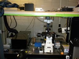
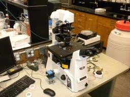
High resolution light microscope. The high resolution light microscope is a multi-mode Nikon TiE microscope that includes widefield fluorescence (10x, 20x,40x and 60x), through-the-objective total internal reflection fluorescence (TIRF) (60x and 100x), differential interference contrast (40x, 60x and 100x) and phase contrast (10x and 20x) microscopy. The stage is heated with an air curtain, but chambers have additional heating for precise temperature control (see below). TIRF illumination includes an argon ion laser with 514/488/457nm lines (150 mW/130 mW/30 mW) and a solid state laser with a 442nm line (45 mW) and shutters to control each laser line separately. Both lines are home-coupled into a single mode fiber that is fed into the TIRF illuminator. Our widefield fluorescence is built around a Prior ProscanII and has excitation/emission/dichroics for 6 different fluorophores. The TiE includes the Perfect Focus System, automated TIRF angle adjustment, and automated Prior shutters and filter wheels for complete control through a computer interface. The two detectors are a low-noise Photometrics CoolSnap HQ2 CCD and Andor iXon EMCCD for high-quality, high-resolution microscopy. The latter is ideal for low signal fluorophores or those prone to photobleaching like GFP and its variants. The microscope and peripherals are driven by mManager, freeware built on ImageJ. Long timelapse microscope. This microscope is a Nikon TiE microsope that includes widefield fluorescence (10x, 20x,40x and 60x), differential interference contrast (40x, 60x and 100x) and phase contrast (10x and 20x) microscopy. The widefield fluorescence is built around a Prior ProscanII and has excitation/emission/dichroics for 2 different fluorophores. The TiE includes the Perfect Focus System and automated Sutter shutters for control through a computer interface. The detector is a CCD from the ImagingSource. The microscope and peripherals are driven by mManager, freeware built on ImageJ. |